
Brown syndrome is a problem with the tendon that attaches to the outside of the eye (superior oblique muscle tendon). In Brown syndrome, this tendon can't move freely. This limits the eye’s normal movements.
The superior oblique muscle is responsible for:
- Pulling the eye toward the midline
- Pulling the eye to look down
- Rotating the eye
The superior oblique muscle tendon attaches to the superior oblique muscle. It moves through a ring of tissue. This is called the tendon sheath.
Many things can limit the normal movement of the muscle tendon through the tendon sheath. When that happens, Brown syndrome occurs.
Brown syndrome is a rare eye disorder. In most cases, a child is born with it (congenital). In very rare cases, it may happen later in life (acquired). Acquired Brown syndrome may be linked to other health conditions. These include injury, inflammatory diseases, problems from eye surgery, and sinus infection. It affects females more often than males.
CAUSES
Despite a substantial amount of research which has helped us better understand Brown Syndrome, the cause is often unknown. Most commonly it is congenital, meaning a child is born with it. Acquired Brown syndrome is uncommon but may be seen following surgery, after trauma, sinus infections or in association with inflammatory diseases. Trauma can cause a Brown Syndrome if a blunt object hits the eye socket in the upper inside corner near the nose. Surgery for the eyelid, frontal sinus, eyeball (retinal detachment) and teeth (dental extraction) have also been linked to acquired Brown syndrome. Inflammation of the tendon-trochlea complex (from adult and juvenile rheumatoid arthritis, systemic lupus erythematosus and sinusitis) can be associated with development of the problem. The actual cause of the restriction of the superior oblique at the trochlea is likely different in all of these situations.
Signs & Symptoms

People with Brown Syndrome have limited eye movement in the affected eye. The ability to move the eyeball toward the center (adduction), or outward from the center (abduction), may be restricted or absent. One eye may appear to be out of alignment with the unaffected eye, especially when looking upward. The symptoms of Brown Syndrome may also include a droopy eyelid (ptosis), widening of the eye (palpebral fissure) when looking upward, crossing of the eyes (strabismus), and/or a backward head tilt. A downward appearance (hypotropia) is usually present in the affected eye when the individual is looking straight ahead (primary position) or in an upward direction. One eye is usually affected, but both eyes (bilateral) may be affected in approximately 10 percent of people with Brown Syndrome.
DIAGNOSED

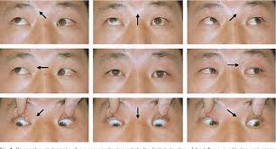
The most common way this is detected in children is that parents will notice the child holds a head turn or chin up head position to compensate for the Brown syndrome. When the child looks up at the parents they will see the eye misalignment that is in figure 2. Often the Brown Syndrome may be so mild that it is not noticed for several years. In these cases the eyes are typically not affected when looking straight ahead and often don’t require treatment. The hallmark sign of Brown syndrome is a decreased ability to look upward and inward with the affected eye(s). In some situations the eyes turn outward (exotropia) when looking up. Occasionally, the affected eye can get “stuck” after looking up or down for long periods of time. When the eye becomes unstuck, a click is often heard and may be accompanied by pain or discomfort. Brown syndrome may be more noticeable in children since they often look upward toward adults.
Treatment:
Treatment will depend on your child's symptoms, age, and general health. It will also depend on how severe the condition is. And on what is causing it.
Close watching often works in mild cases.
More severe cases of Brown syndrome may need surgery. Your child may be more likely to need surgery if:
- The eyes are out of alignment when looking straight ahead
- You child has double vision
- The head position is very abnormal
During this surgery, the eye surgeon may cut the superior oblique muscle tendon and use a device to lengthen it. This may allow the muscle tendon to move normally. The surgery is often successful. But some children need repeat surgery.
Brown syndrome due to other conditions is more likely to go away without surgery. Treating the underlying health condition may help reduce symptoms. For example, someone with Brown syndrome due to lupus might find it helpful to be treated with corticosteroids.
Key points about Brown syndrome in children
- Brown syndrome is a rare problem with a muscle tendon on the outside of the eye. The tendon can't move freely. The eye's normal movement is limited.
- In most cases, this condition is present from birth. In very rare cases, it occurs later in life.
- It often affects only 1 eye. But both eyes can be affected.
- Your child may have trouble looking to the opposite side, and upward, with the affected eye.
- If your child has a mild case, close observation may be advised.
- In more severe cases, children may need surgery.
Next steps
- Tips to help you get the most from a visit to your child’s healthcare provider:
- Know the reason for the visit and what you want to happen.
- Before your visit, write down questions you want answered.
- At the visit, write down the name of a new diagnosis and any new medicines, treatments, or tests. Also write down any new instructions your provider gives you for your child.
- Know why a new medicine or treatment is prescribed and how it will help your child. Also know what the side effects are.
- Ask if your child’s condition can be treated in other ways.
- Know why a test or procedure is recommended and what the results could mean.
- Know what to expect if your child does not take the medicine or have the test or procedure.
- If your child has a follow-up appointment, write down the date, time, and purpose for that visit.
- Know how you can contact your child’s provider after office hours. This is important if your child becomes ill and you have questions or need advice.
Comments
Post a Comment